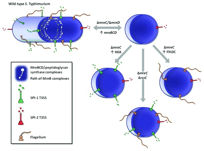
Figure 1. Generation and phenotypic characterization of ΔmreC mutants of S. Typhimurium. Schematic representation of wild-type S. Typhimurium expressing the invasion-associated SPI-1 T3SS, flagella for motility and the SPI-2 T3SS essential for intracellular survival. Wild-type Salmonella cells express 1–2 polar SPI-2 needles and approximately 6–8 flagella and SPI-1 needles, distributed predominantly along the lateral cell wall (Bulmer et al., PLoS Pathog 2012). Putative peptidoglycan synthetic complexes comprising the MreBCD cytoskeletal proteins, cell shape-determinants and peptidoglycan synthetic enzymes, are shown as short dynamic complexes moving perpendicularly to the long-axis of the cell. Upon MreC or MreD depletion S. Typhimurium formed spherical cells, with a loss of SPI-1 T3SS and flagella expression; SPI-2 expression remained active. SPI-1 T3SS and flagella expression were restored upon recovery of either mre, hilA (SPI-1 master regulator) or flhDC (flagella master regulator) expression respectively, or with the inactivation of rcsC, sensor kinase of the Rcs phosphorelay envelope stress response.

An official website of the United States government
Here's how you know
Official websites use .gov
A
.gov website belongs to an official
government organization in the United States.
Secure .gov websites use HTTPS
A lock (
) or https:// means you've safely
connected to the .gov website. Share sensitive
information only on official, secure websites.
