Fig 3.
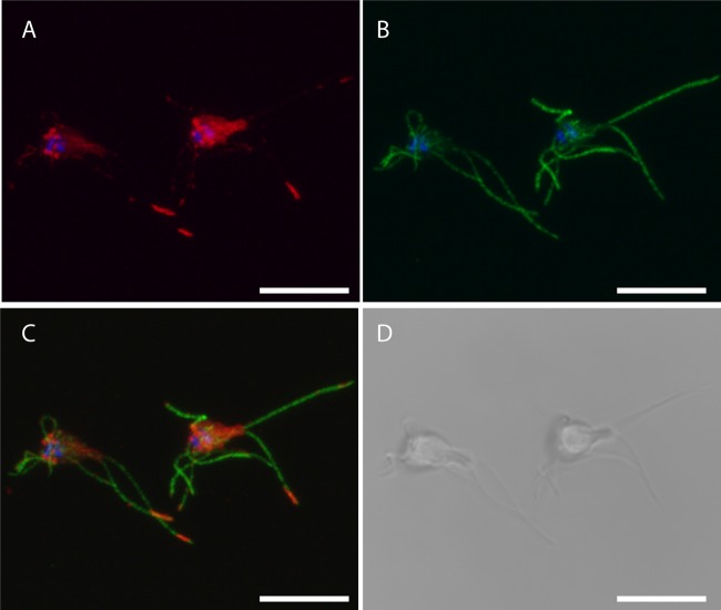
Localization of 3×HA epitope-tagged IFT46 S. salmonicida trophozoites. Transfectants carrying the IFT46-3×HA construct were fixed in 2% PFA and blocked with 2% BSA; they were stained using rabbit anti-HA (1:1,600) and anti-tubulin TAT1 (1:150) and detected using anti-rabbit Alexa Fluor 594 (A594) (1:250) and anti-mouse Alexa Fluor 488 (A488) (1:200), respectively. The cells were mounted in VectaShield medium containing DAPI and viewed using a Zeiss 510 laser scanning confocal microscope. (A) Maximum intensity projection (MIP) of A594 (red) and DAPI (blue) signals; (B) MIP of A488 (green) and DAPI signals; (C) MIP of A488, A594, and DAPI signals; (D) bright-field image from the center of the confocal Z-stack. IFT46-3×HA displays localization to paired cylindrical foci above the nuclei, to sheets that pass through the cell body, and to foci along the flagella sometimes being enriched at the tips. Diffuse staining of the cell body is commonly observed. Scale bars, 10 μm.

