Fig 2.
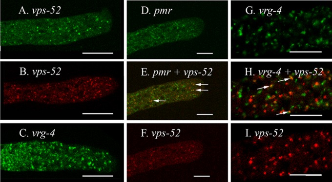
Intracellular location of the PMR, VPS-52, and VRG-4 proteins. The scale bars are 10 μm in panels A to F and 5 μm in panels G to I. (A to C) Cells were transformed with vps-52+::sgfp (A), rfp::vps-52+ (B), and vrg-4+::sgfp (C). (D to F) Shown is a region at the hyphal tip in a heterokaryon formed by the coinoculation of strains transformed with pmr+::sgfp (D) and rfp::vps-52+ (F). (Panels D and F were merged in panel E. (G to I) Shown is a heterokaryon formed by coinoculating strains transformed with vrg-4+::sgfp (G) and rfp::vps-52+ (I). Panels G and I were merged in panel H. Arrows indicate particles with both GFP and RFP. The region shown is approximately 100 μm from the hyphal tip.

