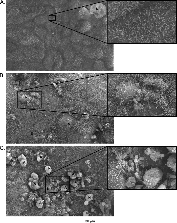
Fig 1.
SEM images of ΔespZ mutant-infected cells reveal larger numbers of dying cells. C2BBE cells were mock treated (A), infected with EPEC (B), or infected with the ΔespZ mutant (C) for 4 h. Selected cells are labeled as follows: A, apoptotic cells; E, effaced cells; N, necrotic cells. Images shown are representative of those collected from cells infected on three separate days. Insets show selected regions at a higher magnification.

