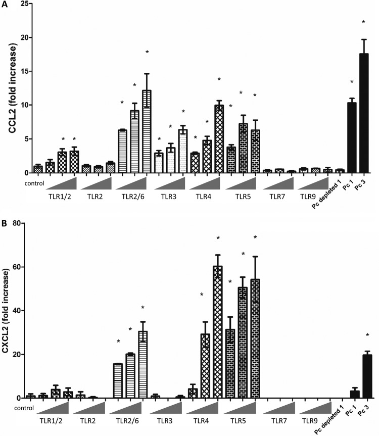
Fig 5.
Primary murine AECs respond to TLR agonists. Primary murine AECs were isolated from C57BL/6 WT mice, and confluent monolayers were stimulated with a panel of TLR ligands for 6 h at increasing concentrations (Table 1): TLR1/2 (Pam3CSK4), TLR2 (heat-killed Listeria monocytogenes), TLR2/6 (FSL1), TLR3 [poly(I·C) and poly(I·C) LMW], TLR4 (E. coli K-12 LPS), TLR5 (S. enterica serovar Typhimurium flagellin), TLR7(ssRNA40), and TLR9 (ODN1826). AECs were also exposed to freshly isolated murine Pneumocystis at cyst-to-AEC ratios of 1:1 (Pc 1) and 3:1 (Pc 3), antibody-depleted Pneumocystis preparations (Pc depleted), and medium alone as negative control (control). CCL2 (A) and CXCL2 (B) levels in the supernatants at 6 h were measured by ELISA. Results are shown as the fold increase relative to the medium-only treatment. Bars represent means ± SEs (n = 3) from a representative experiment that was repeated two times in triplicate. (*, P < 0.05 compared to control).

