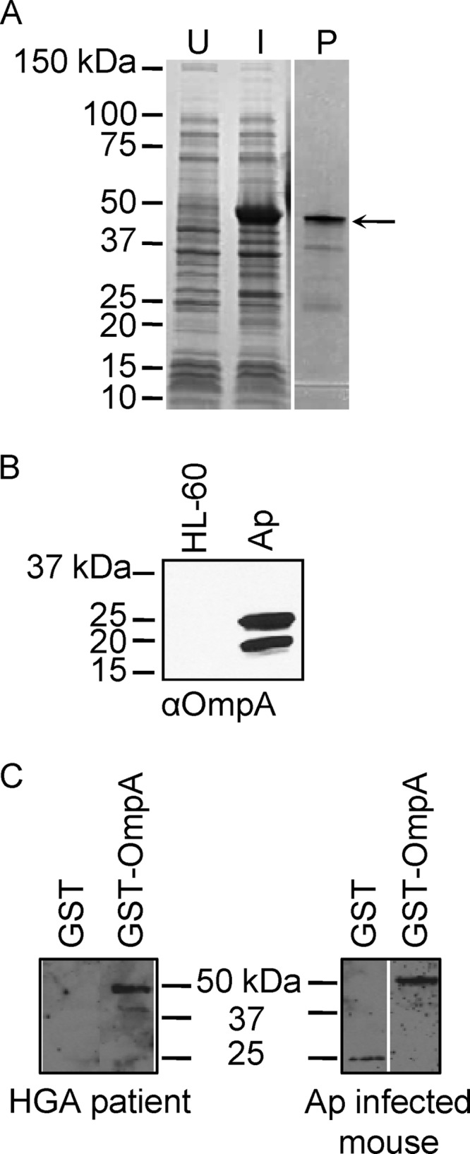
Fig 2.

A. phagocytophilum (Ap) expresses OmpA during in vitro and in vivo infection. (A) Whole-cell lysates of uninduced E. coli (U) and of E. coli induced to express GST-OmpA (I) and GST-OmpA purified by glutathione Sepharose affinity chromatography (P) were separated by SDS-PAGE and stained with Coomassie blue. The arrow denotes the anticipated size for GST-OmpA. (B) Western blot analyses in which mouse anti-OmpA (αOmpA; raised against GST-OmpA) was used to screen whole-cell lysates of uninfected HL-60 cells and A. phagocytophilum organisms derived from infected HL-60 cells. (C) Western blots of GST-OmpA and GST screened with sera from an HGA patient and an experimentally infected mouse.
