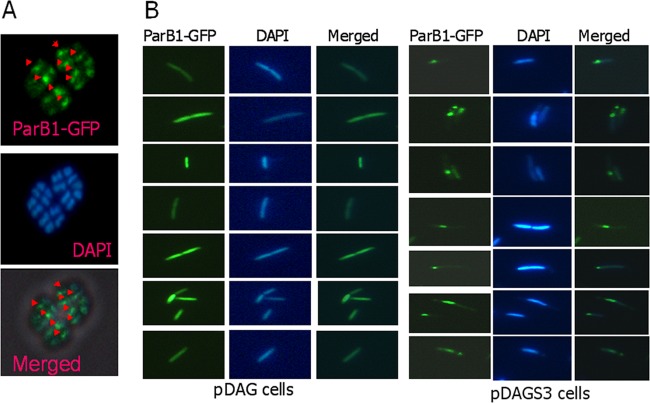
Fig 7.
In vivo interaction of ParB1 with nucleoids containing a segS element(s) in bacteria. D. radiodurans R1 (A) and E. coli (B) cells harboring the pDAG203 vector (pDAG) and pDAGS3 (pDAGS3) were expressed with ParB1-GFP on a plasmid and micrographed for GFP fluorescence (ParB1-GFP), and cells were stained with DAPI. These images were merged to localize the position of the GFP spot on the nucleoid or otherwise, as the case may be.

