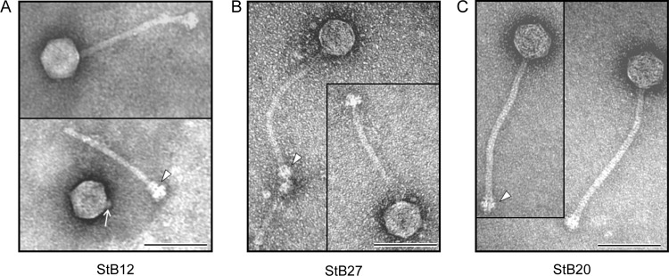
Fig 1.
Morphology of StB20, StB12, and StB27 phages. Transmission electron micrographs show StB12 (A), StB27 (B), and StB20 (C) phage particles. The three phages are characterized by an icosahedral head and a long noncontractile tail. The arrowheads indicate baseplate structures found at the ends of the tails. (A) The arrow indicates the collar structure visible on the virion head of StB12 detached from its tail. Bars, 100 nm.

