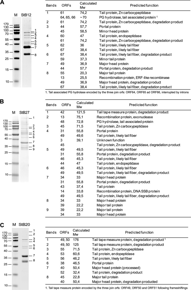
Fig 3.
Structural proteins of STB12, STB27, and STB20 phages. On the left are shown SDS-PAGE gels of proteins extracted from StB12 (A), StB27 (B), and StB20 (C) purified phage particles. The numbers on the left of the gels indicate the sizes of the broad-range mass standard (M). (Right) Identification by LC-MS analysis of proteins extracted from the corresponding SDS-PAGE. For each band, only proteins identified with a Mascot score of ≥120 are considered. Discrepancies between observed and calculated molecular weights (Mw) (in thousands) may be due to specific proteolytic cleavage (see the text) or protein degradation or may be gel migration artifacts.

