
Fig 4.
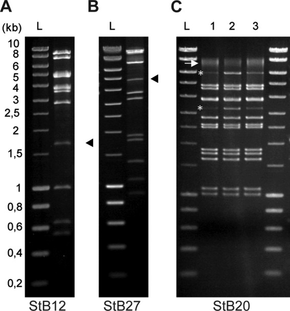
Determination of the DNA-packaging strategy used by StB12, StB20, and StB27 phages. Phage DNA of StB12 (A), StB27 (B), and StB20 (C) was digested by EcoRI, and DNA fragments were visualized on electrophoresis gels stained with ethidium bromide. (A and B) Submolar fragments corresponding to pac fragments observed in the StB12 and StB27 restriction profiles are indicated by arrowheads. (C) (Lane 1) Asterisks indicate the two terminal fragments observed in the StB20 restriction profile. The arrow indicates the two terminal fragments annealed together. (Lanes 2 and 3) Terminal fragments are more visible on the restriction profile obtained after heating and fast cooling of digested DNA (lane 2) than after slow cooling (lane 3), improving the annealing of terminal fragments. Lanes L, size ladders.