
Fig 4.
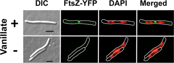
Images obtained using DIC, FtsZ-YFP fluorescence (green), and DAPI fluorescence (red) and a merged image of the last two (green and red) for strain MR2479 grown for 1 h in the presence of 0.5 mM vanillate (top panels) or without vanillate (bottom panels). For the fluorescence images, a white line was drawn bordering the cell boundary. Bars, 5 μm.