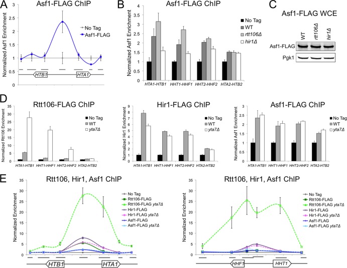
Fig 2.
Asf1 localized with Rtt106 and the HIR complex at histone gene regulatory regions. (A) ChIP analysis of Asf1-FLAG at the HTA1-HTB1 locus (chromosomal coordinates from 913.9 to 916.3 kb). Gray bars indicate the sequences amplified by each qPCR primer set. qPCR values were normalized as described for Fig. 1B. (B) ChIP analysis of Asf1-FLAG at the center of the HTA1-HTB1, HHT1-HHF1, HHT2-HHF2, and HTA2-HTB2 promoters in the indicated mutant strains. WT, wild type. (C) Yeast whole-cell extract (WCE) immunoblotted with anti-FLAG and anti-Pgk1 (loading control) antibodies. (D) ChIP analysis of Rtt106-FLAG (left), Hir1-FLAG (center), and Asf1-FLAG (right) as described for panel B. (E) ChIP analysis of Rtt106-FLAG, Hir1-FLAG, and Asf1-FLAG at the HTA1-HTB1 (left) and HHT1-HHF1 (right, chromosomal coordinates 254.6 to 257.1 kb) loci, as described for panel A.

