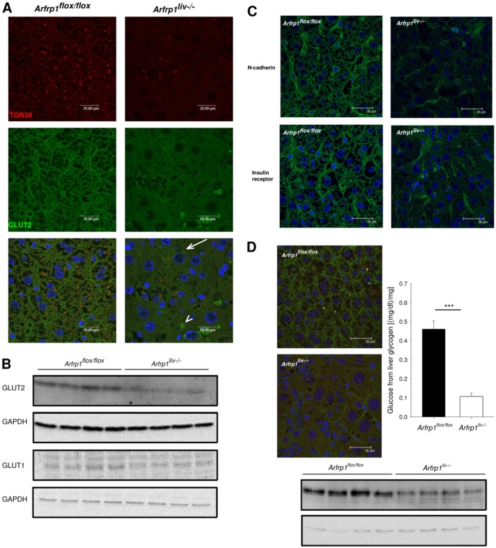
Fig 8.
Reduced plasma membrane localization and protein levels of the glucose transporter GLUT2 in livers of Arfrp1liv−/− mice. (A) Immunohistochemical staining on sections of livers of Arfrp1liv−/− and control mice with an anti-GLUT2 antibody. Cells with successful Arfrp1 deletion were identified by no detectable costaining with an antibody against TGN38. Arrows point to GLUT2 in the plasma membrane of Arfrp1liv−/− mice; arrowheads depict intracellular staining. (B) Western blotting of liver lysates of Arfrp1liv−/− and control mice performed with the indicated antibodies. (C) Normal cell surface localization of N-cadherin and insulin receptor in livers of Arfrp1liv−/− mice as detected by immunohistochemistry. (D) Immunohistochemistry and Western blot of GLUT2 of livers of Arfrp1liv−/− and control mice after 2 weeks of a high-fat diet. The graph shows liver glycogen content in fed Arfrp1liv−/− and control mice.

