Fig 2.
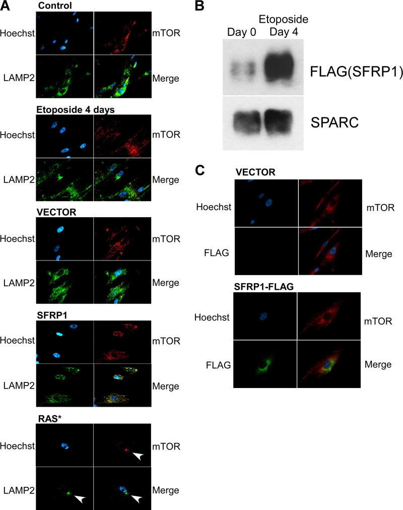
Lack of TASCC formation upon etoposide treatment or SFRP1 expression. (A) Subcellular location of mTOR and LAMP2 upon senescence induced by etoposide, SFRP1, or oncogenic Ras. The formation of TASCC (TOR-autophagy spatial coupling compartment) was assessed by staining for mTOR and LAMP2. Whereas TASCC was readily detectable upon Ras-induced senescence (arrows), TASCC formation was not observed upon etoposide-induced or SFRP1-induced senescence. (B) Etoposide increases the secretion of FLAG-tagged SFRP1. IMR-90 cells were infected with lentiviruses expressing C-terminally FLAG-tagged SFRP1 and were treated with etoposide. The levels of secreted FLAG-tagged SFRP1 were determined by anti-FLAG immunoblotting. (C) Lack of colocalization of FLAG-tagged SFRP1 and mTOR. IMR-90 cells were infected with lentiviruses expressing C-terminally FLAG-tagged SFRP1 or empty vector and were treated with etoposide. Four days later, the cells were stained for mTOR and FLAG.

