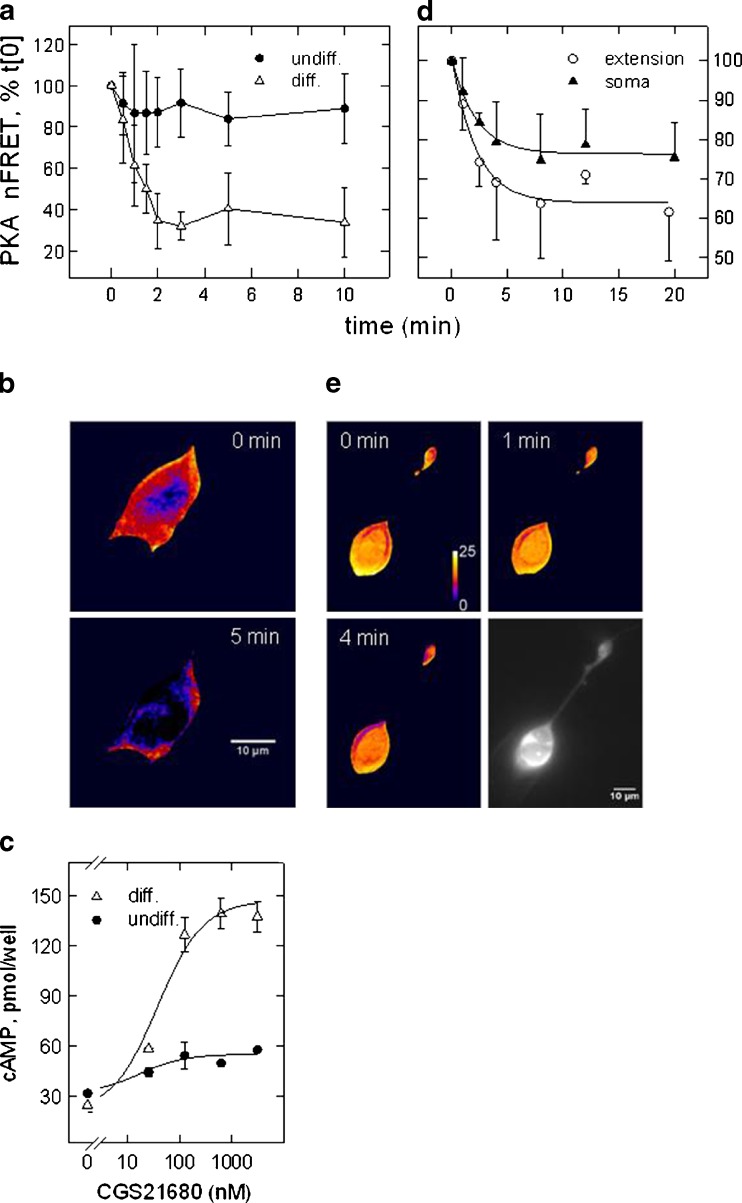
Fig. 1.
A2A-receptor-mediated activation of PKA and stimulation of cAMP formation in SH cells. FRET was recorded of the double-fluorescent PKA construct at various time points following addition of the A2A-receptor agonist CGS21680 (0.5 μM). FRET values were normalised to the fluorescence intensity of donor and acceptor constructs. Plotted are mean values of FRET efficiency (NFRET means ± SD) expressed as percent of the value recorded before addition of the receptor agonist (set 100 %); the data points are connected by a spline curve for graphic purpose. In (a), the experiment was carried out in the presence Ro20-1724 (100 μM) to inhibit PDE activity. Shown are the means of six determinations obtained in differentiated (open triangles) and undifferentiated (filled circles) SH cells. In (d), experiments were performed without addition of Ro20-1724 (n = 4). b, e Images of differentiated cells depicted in false-colour representation of FRET efficiency before and after the addition of the receptor agonist; red to blue transition signifies decreasing FRET efficiency. Images in (b) are representative of the time course depicted in (a) (times 0 and 5 min), images in (e) for the time course in (d) (times 0, 1 and 4 min). Image in grey tones (scale bar = 10 μm) depicts CFP (FRET donor) fluorescence to demonstrate that soma and a neurite extension are from the same cell. FRET-efficiency values for the PKA construct were in the range of 0.25–0.45. c Agonist-dependent formation of cAMP in the presence of Ro20-1724 in differentiated (filled triangles) and undifferentiated (filled circles) SH cells. Cyclic AMP was quantified in diluted cell lysates by ELISA

