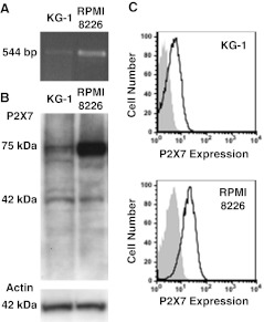
Fig. 1.
KG-1 cells express P2X7. a RNA isolated from KG-1 and RPMI 8226 cells was amplified by RT-PCR using primers to P2X7 and products examined by agarose gel electrophoresis. b Whole cell lysates from KG-1 and RPMI 8226 cells were separated by SDS-PAGE, transferred to nitrocellulose membrane and probed with anti-P2X7 (top panel) or anti-actin (bottom panel) pAb. c KG-1 and RPMI 8226 cells were labeled with Alexa Fluor 647-conjugated P2X7 (solid line) or isotype control (shaded) mAb, and the relative cell-surface P2X7 expression measured by flow cytometry. Representative results from (a,b) two or c three experiments shown

