
Fig 5.
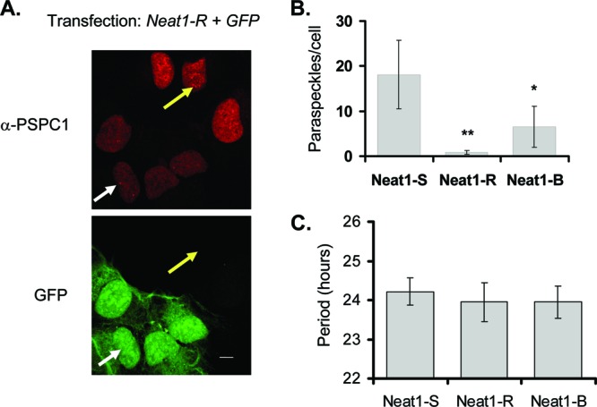
(A) Immunofluorescence from cells transfected with a plasmid expressing green fluorescent protein (GFP) and an RNAi construct targeting Neat1 (Neat-R). White arrow, paraspeckle in transfected cell; yellow arrow, paraspeckle in untransfected cell; α, anti. Scale bar, 10 μm. (B) Quantification (±standard deviation) of paraspeckles per cell for two different RNAi constructs (R and B), as well as a scrambled hairpin (S), quantified by immunostaining as described for panel A (n = 12 cells for Neat1-R, 24 for Neat1-B, and 18 for Neat1-S). *, P < 0.05; **, P <0.01 (Student t test.) (C) Period length of circadian reporter expression for U2OS cells cotransfected with the hairpins described for panel A and the Bmal1-luc circadian reporter. (n = 6 per sample; no significant differences, as determined by a Student t test).