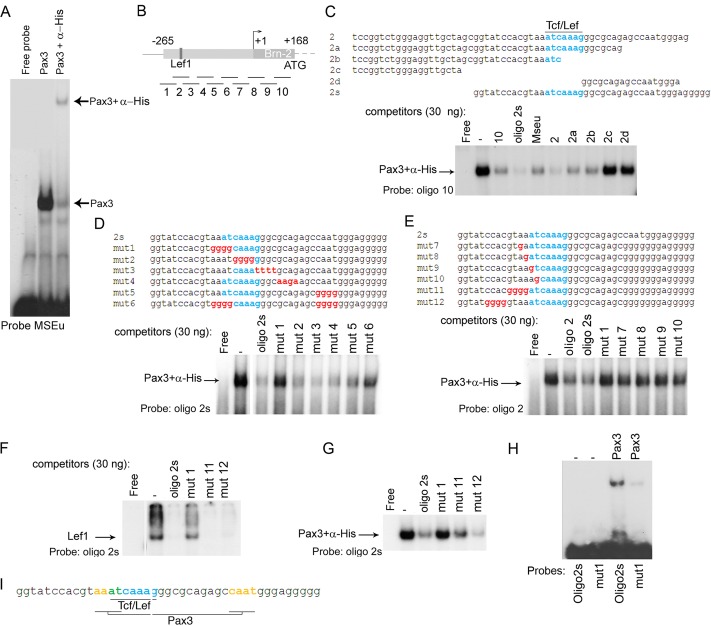
Fig 4.
Pax3 directly binds the Brn-2 promoter. (A) Band shift using the MSEu probe from the Tyrp1 promoter together with bacterially expressed and purified Pax3-PDHD-His tagged with or without α-His antibody. (B) Schematic of the Brn-2 promoter showing the Tcf/Lef1 binding site. The lines under the Brn-2 promoter represent the locations of 10 oligonucleotides used as probes or competitors in our band shift assays. (C) Band shift assay using an excess of probe (oligonucleotide 10) together with bacterially expressed and purified Pax3-PDHD-His. Thirty nanograms of the indicated competitors corresponding to a region of the proximal promoter of Brn-2 was used. The described Tcf/Lef1 binding site is highlighted in blue. (D) Binding of Pax3 to the probe oligonucleotide 2s was challenged using 30 ng of the indicated competitors. Blue, Lef1 binding site; red, mutated bases of the potential Pax3 binding site. (E) Binding of Pax3 to the probe oligonucleotide 2 was challenged using 30 ng of the indicated competitors. (F) Band shift using bacterially purified GST-Lef1 together with oligonucleotide 2s and 30 ng of unlabeled oligonucleotide 2s corresponding to the probe and mutant 1, 11, and 12 as competitors. (G) Binding of Pax3 to the probe oligonucleotide 2 was challenged using 30 ng of the indicated competitors. (H) Binding of Pax3 to WT oligonucleotide 2s or Mut1 probes as indicated. (I) Scheme representing the Pax3 binding sites (yellow) adjacent to Lef1 (blue) on the Brn-2 promoter. The bases shared by both Pax3 and Lef1 are represented in green.

