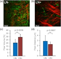
Fig. 3.
Primary breast tumors from lymph node positive () patients contained denser Col1 fibers than primary tumors from patients. Shown are representative SHG Col1 fiber (red) images from an patient (a) and an patient (b). Nuclei were counterstained with Hoechst (green). Each image size is 339 by 339 μm; one -plane is displayed. The scale bars represent 50 μm. patients () displayed significantly () decreased fiber volume (c) and a trend () toward increased inter-fiber distance (d) compared to patients (). Values are .

