Fig 5.
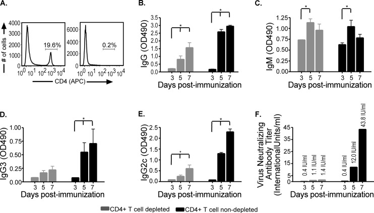
Antibody responses in rRV-ΔM-immunized mice depleted of CD4+ T cells. C57BL/6 mice were depleted of CD4+ T cells using anti-CD4 antibody (GK1.5) as described in Materials and Methods and then immunized intramuscularly with 106 FFU/mouse rRV-ΔM. Sera from immunized mice were collected on the indicated days. Spleens from CD4+ T cell-depleted and wild-type mice were collected throughout the sample period and tested for CD4+ T cell depletion. (A) Depletion of CD4+ T cell-depleted mice (right panel) at all time points, including day 7 postimmunization, was determined to be 95% to 99% compared to nondepleted mice (left panel) as shown by representative analysis of CD4 T cells. (B through E) On the indicated times postimmunization, RV glycoprotein (G)-specific IgG (B), IgM (C), IgG3 (D), and IgG2c (E) antibodies were determined by ELISA. OD490, optical density at 490 nm. (F) Virus-neutralizing antibodies were also determined as described in the legend to Fig. 4. While wild-type mice had higher VNA titers than CD4+ T cell-depleted mice, the latter showed antibody titers indicative of a satisfactory immunization (i.e., >0.5 IU/ml) as early as 4 days postimmunization in the absence of CD4+ T cell help. IgG (of all subclasses tested) increased over time in CD4+ T cell-depleted mice, and IgM antibodies were also detected. n = 5/group of CD4+ T cell-depleted mice; n = 4/group of wild-type mice; to compare two groups of data, we used an unpaired, two-tailed t test; *, P < 0.05.

