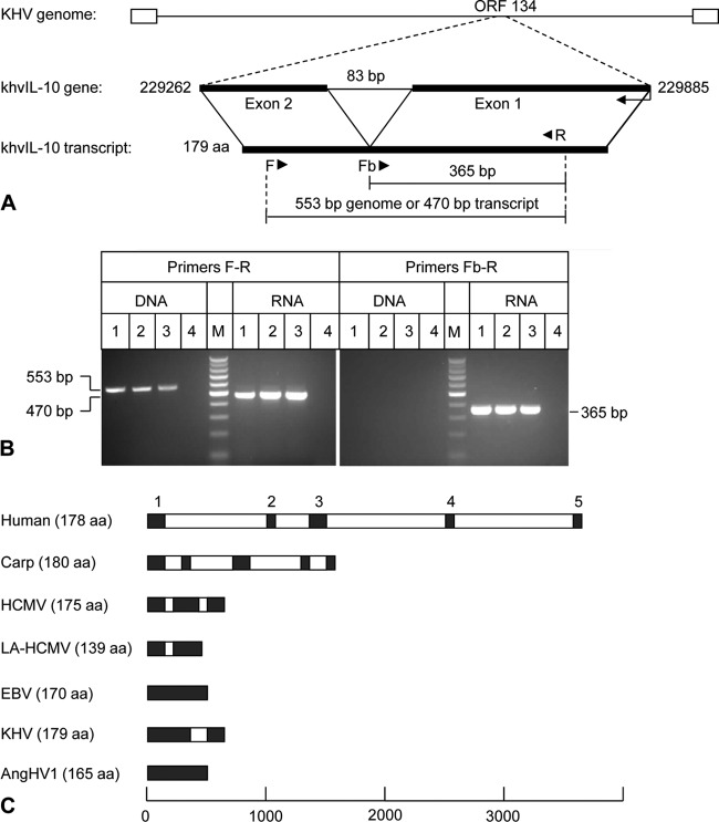
Fig 2.
PCR and RT-PCR analysis of the khvIL-10 gene and comparison of splicing patterns of host and viral IL-10 genes. (A) Predicted structure of KHV ORF134 and locations of primer binding sites. (B) Agarose gel electrophoresis of products amplified from DNA and total RNA extracted from KHV-infected cells using PCR primer sets F-R and Fb-R. Lanes 1, KHV U.S. strain F9850; lanes 2, United Kingdom strain G406; lanes 3, Indonesian strain C07; lanes 4, mock infected; lanes M, 100-bp DNA ladder. (C) Structures of host IL-10 and viral IL-10 genes. Introns (white boxes) and exons (black boxes) are indicated, and exon numbers for human IL-10 are provided.

