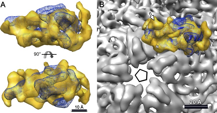
Fig 7.
Orientation of P7 in the procapsid. (A) Crystal structure of ϕ12 P7 (blue ribbon) (PDB ID 2Q82) (6) fitted into the P7 density map (yellow) from the P1247-P124 difference map. The fitting, which involved a global search over all orientations, is described in Materials and Methods. (B) In this setting, the C- and N-terminal helices of P7 both lie close to the 5-fold vertex of the procapsid. Both P7 termini were implicated in RNA binding and could thus interact with packaged RNA in this orientation. Additionally, the EM map rendered at a lower threshold shows an unassigned density near the vertex (arrow) that may correspond to a part of the C-terminal region of P7 that is missing from the crystal structure.

