
Fig 1.
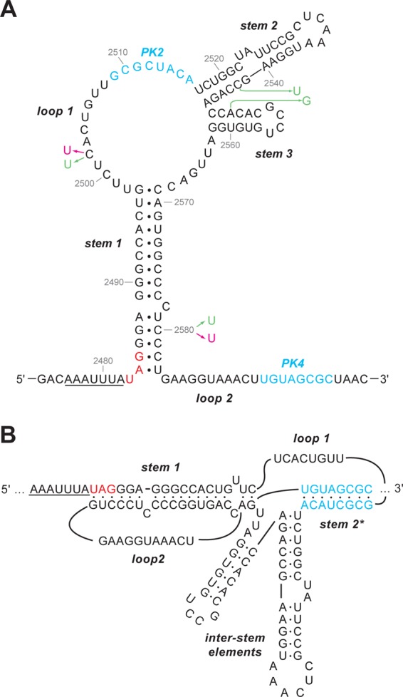
Secondary structure of the Rous sarcoma virus frameshift site (33). (A) The slippery sequence is underlined, and the gag stop codon is shown in red. The complementary PK2 and PK4 regions involved in forming the pseudoknot are in blue. The sequence shown is that of the Prague C strain (PrC) (43). Nucleotides that differ in ALV are in pink, and those that differ in the pRCAS proviral clone, which is based on the Schmidt-Ruppin strain (subgroup A), are in green. (B) An alternative view shows the pseudoknot formation more clearly, with stems 2 and 3 as interstem elements. In this representation, base pairing of PK2 and PK4 leads to the formation of pseudoknot stem 2*, which is marked with an asterisk to distinguish it from the unrelated stem 2 in the representation shown in panel A.