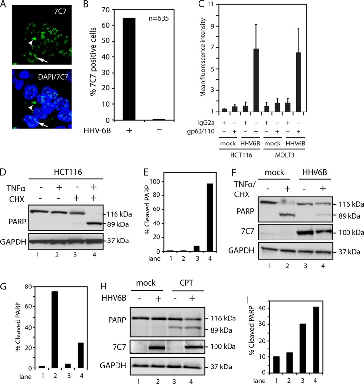
Fig 1.
HHV-6B infection inhibits TNF-α-induced apoptosis. (A) Confocal microscopy of HCT116 cells infected with HHV-6B for 48 h and stained with 7C7, an HHV-6A/B-specific antibody. The arrows point to nuclear viral proteins, and the arrowheads point to cytoplasmic proteins. DAPI, 4′,6-diamidino-2-phenylindole. (B) Quantification of 7C7-positive cells from confocal microscopy of HCT116 cells mock treated or infected with HHV-6B for 48 h and stained with 7C7 antibody. (C) HCT116 and MOLT3 cells were infected with HHV-6B for 48 h and stained with isotype- or gp60/110-specific antibodies, followed by flow cytometry analyses. The graph represents an average of four independent experiments. The error bars indicate standard deviations (SD). (D) Western blot analyses with antibodies against PARP or GAPDH (glyceraldehyde-3-phosphate dehydrogenase) (loading control). HCT116 cells were treated with TNF-α (25 ng/ml), CHX (10 μM), or a combination of both for 4 h, followed by Western blot analyses with antibodies against PARP or GAPDH. (E) Quantifications of cleaved PARP from panel D. (F) Western blot analysis of lysates from mock-treated or HHV-6B-infected (48 h) HCT116 cells, followed by treatment with TNF-α and CHX for 4 h. The Western blots were probed with antibodies against PARP or GAPDH. (G) Quantifications of cleaved PARP from panel F. (H) Western blot analyses of lysates from mock-treated or HHV-6B-infected (48 h) HCT116 cells, followed by treatment with CPT for 18 h. The Western blots were probed with antibodies against PARP or GAPDH. (I) Quantifications of cleaved PARP from panel H. Representatives of at least two experiments are shown.

