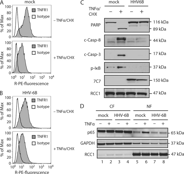
Fig 2.
HHV-6B infection inhibits proinflammatory and apoptotic TNFR1 signaling pathways. (A to C) HCT116 cells were mock treated or HHV-6B infected (48 h) and then incubated in the presence or absence of TNF-α (25 ng/ml) and CHX (10 μM) for 4 h, followed by flow cytometry analyses with TNFR1- and isotype-specific antibodies (A and B) or Western blot analyses with antibodies against PARP, cleaved caspase 8 (c-Casp-8), cleaved caspase 3 (c-Casp-3), p-IκB, 7C7 (HHV-6B infection control), and RCC1 (loading control) (C). (D) HCT116 cells were mock treated or infected with HHV-6B (48 h) and then incubated in the presence or absence of TNF-α (25 ng/ml) for 30 min. Shown are Western blot analyses of cytoplasmic (CF) and nuclear (NF) fractions with antibodies against p65 or the loading controls GAPDH and RCC1. Representatives of at least two experiments are shown.

