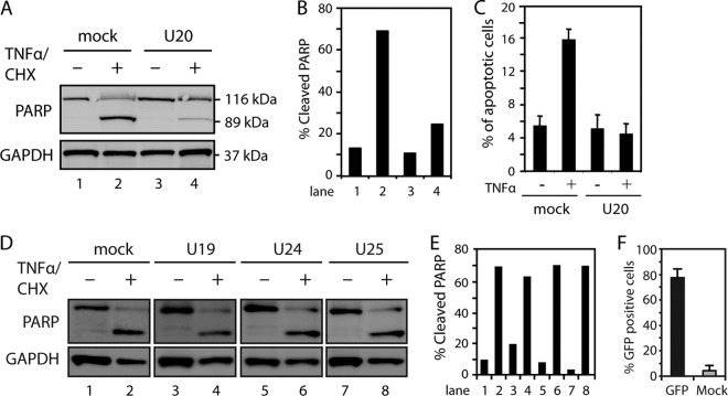
Fig 3.
HHV-6B-encoded protein U20 inhibits TNF-α-induced apoptosis. (A) HCT116 cells were transfected with plasmid alone (mock) or a plasmid carrying U20 and left untreated or treated with TNF-α (25 ng/ml) and CHX (10 μM) for 4 h, followed by Western blot analysis with antibodies against PARP or GAPDH (loading control). (B) Quantification of cleaved PARP from panel A. (C) HCT116 cells were transfected with plasmid alone or a plasmid carrying U20 and treated with TNF-α and CHX for 4 h, followed by 7-AAD staining and flow cytometry analysis. The average percentages of apoptotic cells from three experiments are shown. The error bars indicate SD. (D) HCT116 cells were transfected with plasmid alone or a plasmid carrying U19, U24, or U25 and left untreated or treated with TNF-α and CHX for 4 h, followed by Western blot analysis with antibodies against PARP or GAPDH (loading control). (E) Quantification of cleaved PARP from panel D. Representatives of at least two experiments are shown. (F) HCT116 cells were transfected with plasmid alone or a plasmid encoding GFP for 24 h, followed by flow cytometry analyses. An average of four independent experiments is shown. The error bars indicate SD.

