Fig 4.
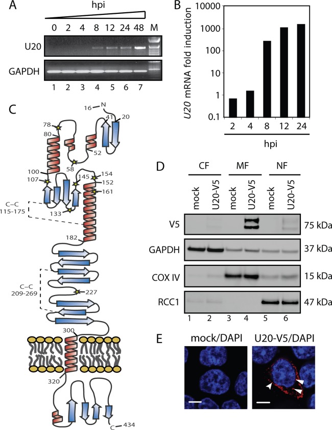
U20 is a 75-kDa membrane-associated protein. (A) PCR with primers spanning the U20 ORF on cDNA from MOLT3 cells infected with HHV-6B for 0, 2, 4, 8, 12, 24, or 48 h. GAPDH served as a control. M, base pair marker. (B) Real-time PCR with U20 ORF-specific primers on cDNA from HCT116 cells infected with HHV-6B for 0, 2, 4, 8, 12, or 24 h. The amount of U20 was quantified relative to β-actin and is presented as fold induction relative the 0-h measurement. (C) Hypothetical model of U20. Shown are signal peptides (aa 1 to 15), the immunoglobulin-like domain (aa 182 to 288), and the transmembrane α-helix (aa 300 to 320). Predicted α-helices are shown in red, and predicted β-sheets are shown in blue. Predicted C-C bridges between Cys-115 and Cys-175 and between Cys-209 and Cys 269 are indicated by broken lines. (D) HCT116 cells were transfected with plasmid alone or a plasmid carrying N-terminally V5-tagged U20. Shown are Western blot analyses of cytoplasmic (CF), membrane (MF), and nuclear (NF) fractions with antibodies against V5 epitope or the loading controls GAPDH, COX IV, and RCC1. (E) HCT116 cells were transfected with plasmid alone or a plasmid carrying N-terminally V5-tagged U20 and subjected to confocal microscopy with an antibody against the V5 epitope (red) and DAPI (blue). Scale bars, 10 μm. The curved arrowhead points to U20 associated with the surface membrane, and the full arrowheads point to U20 associated with intracellular membranes.

