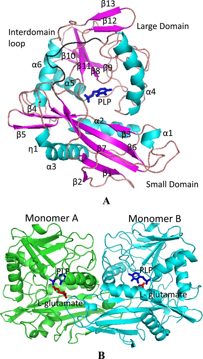
Fig 3.

Crystal structures of DrBCAT and molecular packing. (A) The PLP-bound monomer structure of DrBCAT is shown with six α-helices (turquoise) and 13 β-strands (magenta). The overall structure comprises small and large domains, connected to the interdomain loop (Val172 to Phe185; shown in black) located on the molecular surface. The cofactor PLP (dark blue stick) is located at the bottom of the active site between the two domains. (B) The DrBCAT–l-glutamate dimer (space group P21) is shown from a direction nearly perpendicular to the 2-fold axis. The PLP (dark blue stick) and l-glutamate (red stick) are located at the bottom of the active site.
