
Fig 4.
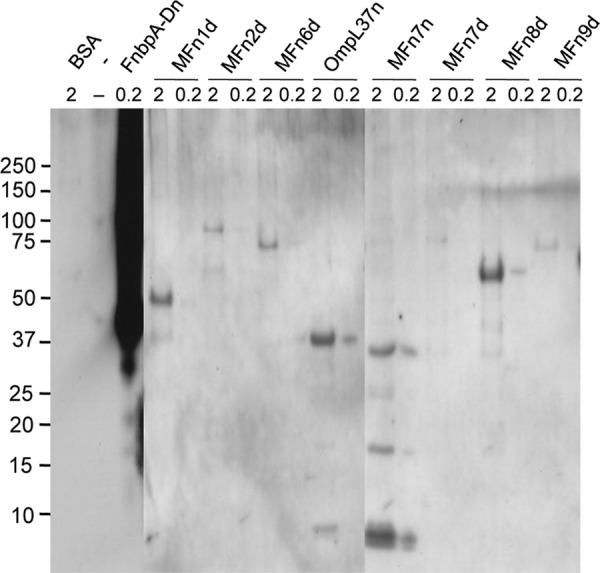
Ligand affinity blot (far-Western) of rMFn proteins. Recombinant proteins were separated by gel electrophoresis, blotted onto PVDF membranes, and probed with 10 μg of human plasma fibronectin/ml. Recombinant OmpL37 (80) and S. aureus FnbpA D repeats (19, 102) were included as positive controls, and BSA was included as a negative control. An “n” or a “d” denote purification of recombinant proteins either under native or denaturing conditions, respectively, as described in Materials and Methods, as well as in previous reports (19, 80). The quantity of protein per lane is indicated (2 or 0.2 μg). The positions of molecular mass standards (in kilodaltons) are indicated on the left.