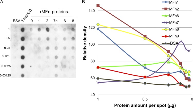
Fig 5.
Semiquantitative dot blot. The data are representative of four independent dot blot experiments. (A) Recombinant proteins were transferred to a 0.45-μm-pore-size nitrocellulose membrane by microfiltration and probed with 10 μg of human plasma fibronectin/ml. The micrograms of protein per spot are indicated on the left. BSA was included as a negative control, and the S. aureus FnbpA-D repeat protein (19, 102) was included as a positive control. Only FnbpA-D (19) and MFn7 (see Materials and Methods) were purified under native conditions. Duplicate spots were included in each experiment. (B) The intensities of each spot were analyzed by ImageJ software by subtracting the background and measuring the mean density of the pixels in each spot. The mean values of duplicate spots are displayed.

