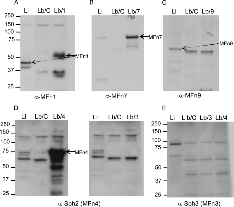
Fig 8.
Expression of MFn proteins by L. biflexa transformants. Whole-cell lysates of 108 leptospires per lane were separated by gel electrophoresis, blotted onto PVDF membrane, and probed with rabbit immune sera recognizing MFn1 (A), MFn7 (B), MFn9 (C), MFn4 (Sph2) (D), and MFn3 (Sph3) (E). Lanes: Li, L. interrogans serovar Copenhageni strain Fiocruz L1-130; Lb/C, L. biflexa serovar Patoc strain Patoc I transformed with an empty pRAT575 vector, used as a control; Lb/1, Lb/3, Lb/4, Lb/7, and Lb/9, L. biflexa Patoc I transformed with pRAT575 vector constructs containing mfn1, mfn3, mfn4, mfn7, and mfn9 genes, respectively. The positions of molecular mass standards (in kilodaltons) are indicated on the left.

