Fig 1.
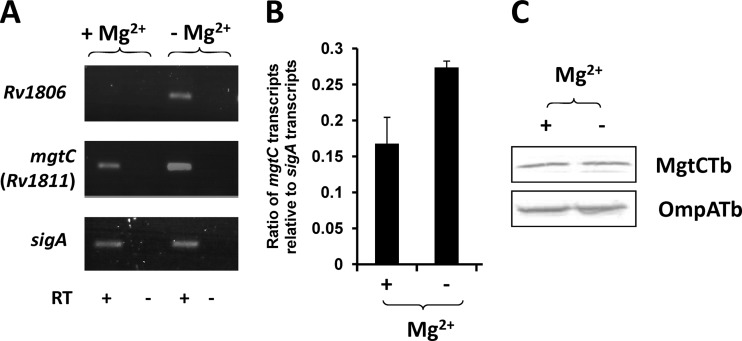
Expression of M. tuberculosis mgtC transcripts and the M. tuberculosis MgtC protein under high-Mg2+ and low-Mg2+ conditions. (A) RT-PCR experiment with RNA isolated from M. tuberculosis grown in high or low Mg2+. Reverse transcriptase was omitted in two lanes (as indicated by “-”) to verify the absence of genomic DNA contamination in the RNA sample. (B) Quantification of M. tuberculosis mgtC RNA by a q-RT-PCR experiment using RNA isolated from M. tuberculosis grown in high or low Mg2+. The sigma factor sigA was used as an internal standard. Results are expressed as means of the mgtC/sigA ratio ± standard deviations from three independent experiments. (C) Bacterial lysates equivalent to 40 μg of total proteins were separated by 15% SDS-PAGE. For the detection of M. tuberculosis MgtC, the membrane was incubated with rabbit anti-M. tuberculosis MgtC antibodies at a 1:500 dilution, washed, and probed with anti-rabbit antibodies conjugated to alkaline phosphatase used at a 1:7,000 dilution. The loading control was performed by probing the same amount of fractions with rabbit anti-OmpATb73–326 antibodies at a 1:6,000 dilution.

