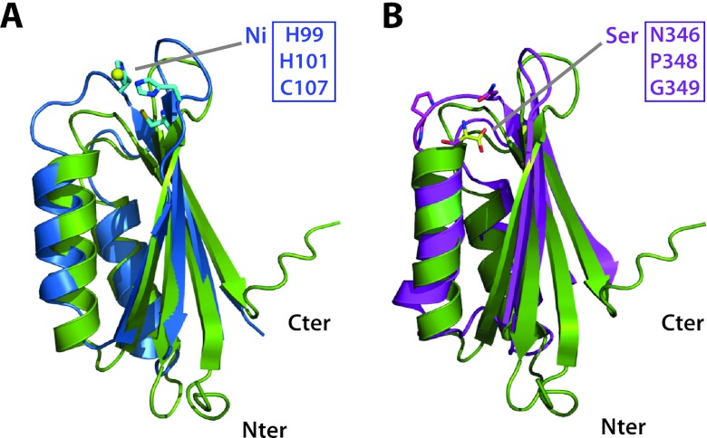
Fig 4.
Superposition M. tuberculosis MgtC-Cter on other ACT domains. (A) Superposition of the ACT domains of M. tuberculosis MgtC (PDB 2LQJ; in green ribbon) and of nickel-bound NikR from Helicobacter pylori (PDB 3LGH; blue ribbon). The side chains involved in metal binding are shown as sticks. (B) Superposition of the ACT domains of M. tuberculosis MgtC (in green ribbon) and of serine-bound SerA from Escherichia coli (PDB 1PSD; pink ribbon). Three residues building up the amino acid binding site and the bound serine are shown as sticks. These residues are conserved in the amino acid binding site of ACT domains but are absent in members of the MgtC family and NikR (see Fig. 3). Two additional residues (H344 and N364; not as well conserved in M. tuberculosis MgtC) contacting the serine ligand through hydrogen-bound are not shown for clarity. The figure was prepared using the program Pymol (http://www.pymol.org).

