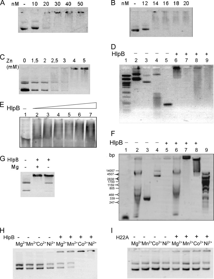
Fig 4.
Gel retardation experiments (0.8% agarose, except for panel E, for which a 5% to 10% native polyacrylamide gradient gel was employed) using purified HlpB. (A and B) Plasmid DNA (0.5 μg/6 nM) was incubated with different amounts of HlpB as indicated above the lanes in the presence of 5 mM ZnCl2. (C) Plasmid VK1 (0.5 μg/6 nM) was incubated with 100 nM HlpB and different concentrations of Zn2+ as indicated. (D) Different DNA molecules were incubated with 5 mM ZnCl2 and without (lanes 2 to 5) or with (lanes 6 to 9) 200 nM HlpB. Lane 1, marker DNA; lanes 2 and 6, pVK1 (0.5 μg); lanes 3 and 7, pQE60 (0.5 μg); lanes 4 and 8, B. subtilis chromosomal DNA (0.5 μg); lanes 5 and 9, 500-bp PCR fragment (0.5 μg). Note that the shifted band is not in but is underneath the wells of the gel. (E) A 70-bp dsDNA oligonucleotide (3.2 μg) was incubated with increasing amounts of HlpB. Lane 1, no HlpB; lane 2, 1.5 μg HlpB; lane 3, 2.5 μg HlpB; lane 4, 3 μg HlpB; lane 5, 3.5 μg HlpB; lane 6, 4 μg HlpB; lane 7, 4.5 μg HlpB. (F) Different DNA molecules were incubated without (lanes 1 to 4) or with (lanes 5 to 8) 200 nM HlpB and 0.5 mM Zn2+. Lane 9, PstI-digested DNA; lanes 4 and 8, plasmid DNA (100 ng); lanes 3 and 7, 500-bp PCR fragment (0.5 μg); lanes 2 and 6, chromosomal Bacillus subtilis DNA (0.5 μg); lanes 1 and 5, ssDNA oligonucleotide (0.5 μg). (G) Plasmid DNA was incubated with 2 mM ZnCl2, without or with 100 nM HlpB, and without or with 5 mM MgCl2 as indicated. (H) Plasmid DNA was incubated with or without 100 nM HlpB and with different metal ions (5 mM) as indicated above the lanes. (I) Plasmid DNA was incubated with or without 100 nM H22A mutant HlpB and with different metal ions (5 mM) as indicated above the lanes.

