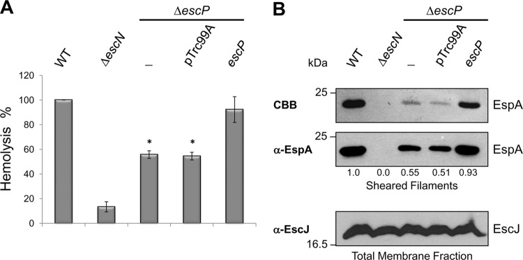
Fig 4.
An escP mutant EPEC strain shows reduced hemolytic activity and assembles fewer EspA filaments. (A) Hemolytic capabilities of WT EPEC strain, the ΔescN mutant strain, the ΔescP mutant strain (−), the ΔescP mutant strain with the empty vector (pTrc99A), and the ΔescP mutant strain expressing the plasmid pJTo16 (escP). Hemoglobin released into the supernatant was measured by determining OD450 after incubation of EPEC strains with human RBCs. Standard deviations of three independent experiments are shown. Asterisks indicate significant differences from WT EPEC. *, P < 0.0001, as determined by Student's t test. (B) EspA filament sheared from WT EPEC, the ΔescN mutant strain, the ΔescP mutant strain (−), the ΔescP mutant strain with the empty vector (pTrc99A), and the ΔescP mutant strain expressing plasmid pJTo16 (escP). EspA protein was visualized by Coomassie brilliant blue (CBB)-stained SDS–12.5% PAGE (upper panel) and by immunoblotting with anti-EspA antibody (middle panel). Densitometry analysis of EspA bands in the Western blot assay was performed with the Scion Image software (Scion Corporation). Membrane fractions of sheared bacterial cells were analyzed by immunoblotting with anti-EscJ antibody (lower panel). Molecular masses of protein standards are indicated on the left of each panel.

