Fig 2.
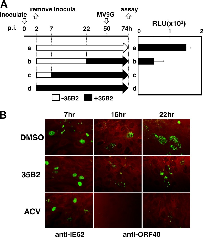
(A) “Time of addition” experiment. MeWo cells were inoculated with cell-free VZV (V-Oka) at the time point 0 h. The inocula were removed at 2 h, and fresh media without (a, b, and c) and with (d) 35B2 were added to the cells. 35B2 was added to the cells at the time points 7 h (c) and 22 h (b). At 50 h, MV9G VZV reporter cells were added and cultured for an additional 24 h. Luciferase activities were expressed in terms of relative light units (RLU). (B) HEL cells were infected with cell-free VZV (V-Oka). Two hours later, the inocula were removed. The cells were cultured in the presence of DMSO, 20 μM 35B2, and 20 μg/ml ACV for the indicated durations and fixed with acetone. The antibodies used for IFA are indicated. Evans blue was used for counterstaining.

