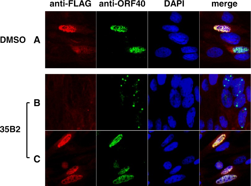
Fig 8.
Effect of overexpression of scaffold proteins on localization of the VZV MCP in infected cells. GPL cells were plated in media containing DMSO (A) or 20 μM 35B2 (B and C; same well) and transfected with pCMV-ORF33F expressing FLAG-tagged ORF33. One day after transfection, the cells were infected with V-Oka. Two days after infection, the cells were fixed with acetone. The FLAG-tagged ORF33 protein and ORF40 protein were visualized by IFA using anti-FLAG tag monoclonal antibody and rabbit anti-ORF40 polyclonal antibody as primary antibodies, Alexa Fluor 594-conjugated anti-mouse IgG Fab′ and FITC-conjugated anti-rabbit IgG Fab′ as secondary antibodies, and DAPI for counterstaining. Alexa Fluor 594, FITC, and DAPI signals are shown in red, green, and blue, respectively. There were cells with (C) and without (B) expression of FLAG-tagged ORF33 in the same well, since the transfection efficiency was 30 to 40%.

