Fig 4.
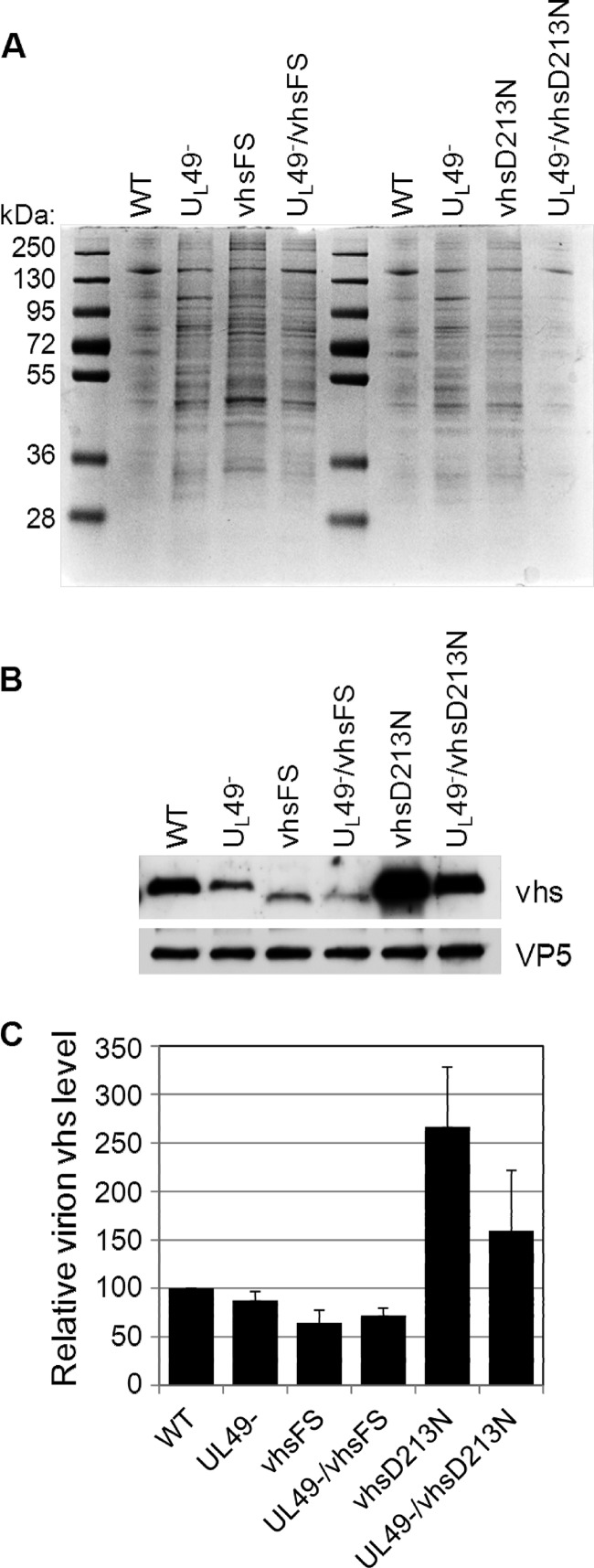
Relative virion vhs levels. Virions were purified from V49 cells infected with the WT, UL49−, vhsFS, UL49−/vhsFS, vhsD213N, or UL49−/vhsD213N virus at an MOI of 0.005 PFU/cell and collected when CPE reached 100%. (A) Proteins present in purified virions were separated by SDS-PAGE and stained with Coomassie brilliant blue. Results are representative of three independent experiments. (B) Virion proteins were also examined by immunoblotting using an antibody specific to VP5 followed by blot stripping and reprobing with an antibody specific to vhs. The vhsFS mutation results in early translation termination and, thus, faster migration during SDS-PAGE. Results are representative of three independent experiments. (C) Immunoblot signals were quantified, and the vhs signal on each blot was normalized to the corresponding VP5 signal. Data were compiled from three independent experiments. Values shown are arithmetic mean VP5-normalized vhs levels present in virions relative to those in the WT; error bars represent 1 standard deviation.

