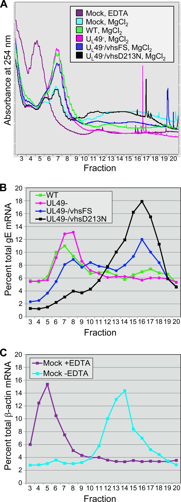
Fig 6.
Analysis of translation efficiency. Postmitochondrial supernatants prepared from Vero cells mock treated or infected at an MOI of 10 PFU/cell for 18 h with the WT, UL49−, UL49−/vhsFS, or UL49−/vhsD213N virus were separated by 10% to 50% sucrose density centrifugation. Mock-treated supernatants were prepared in the presence and absence of EDTA. Gradients were driven, top to bottom, through a UV-Vis continuous flow cell by the peristaltic pump of a gradient fractionator. Gradients were fractionated as they exited the flow cell. (A) UV absorbance profiles at 254 nm of fractions 3 to 20 from each gradient. The top of the gradient is to the left, and the bottom is to the right. Sharp spikes in the UV absorbance toward the bottom of the gradient were caused by air bubbles that form in the peristaltic pump tubing in denser regions of the gradient. Similar results were observed in three independent experiments. (B and C) Distribution of mRNAs across gradient fractions collected following UV absorbance measurements. RNA was extracted from gradient fractions and analyzed by Northern blotting using probes specific to gE (B) or β-actin (C). Bands were quantified on a phosphorimager, and the signal in each fraction in a gradient was calculated as a percentage of the signal across all fractions in that gradient to determine the distribution of the mRNAs across the gradients. The graphs represent one of two independent, comparable experiments.

