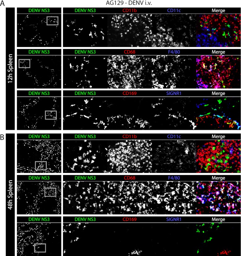
Fig 5.
In the spleen, DENV first infects macrophages of the marginal zone and later infects macrophages of the red pulp. (A) Representative immunohistochemical staining of spleen sections from AG129 mice 12 h after infection (5 × 10⁸ GE of the DENV2 strain E124/128-IC i.v. on day 0). Tissue sections were stained for DENV NS3 (green) and CD11b and CD11c (red and blue, respectively, top row), CD68 and F4/80 (red and blue, respectively, middle row), and CD169 and SIGN-R1 (red and blue, respectively, bottom row). Low-magnification (×5) images are shown on the left with DENV NS3 alone, while each individual channel and the merged image are shown on the right. Gray boxes on the DENV NS3 panels depict the areas of enlargement shown on the right. The results are representative of 3 mice. (B) Representative immunohistochemical staining of spleen sections from AG129 mice 48 h after infection (5 × 10⁸ GE of the DENV2 strain E124/128-IC i.v. on day 0). Sections are stained as described for panel A. The results are representative of 3 mice.

