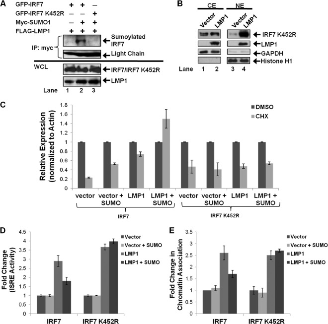
Fig 8.
LMP1-induced sumoylation of IRF7 occurs at IRF7 lysine 452. 293T cells were transfected as indicated. (A) Cell lysates were collected 48 h posttransfection. Denaturing immunoprecipitations were performed with c-Myc antibodies. Western blot analyses were performed on the immunoprecipitants and whole-cell lysates to determine IRF7, sumoylated IRF7, IRF7 K452R, sumoylated IRF7 K452R, and LMP1 expression. (B) Cytoplasmic (CE) and nuclear (NE) extracts were collected, and Western blot analyses were used to detect IRF7 K452R and LMP1. Histone H1 and GAPDH were used as loading controls. (C) Eighteen hours before harvesting, cells were treated with DMSO or CHX (75 μg/ml). Cell lysates were harvested 48 h posttransfection and immunoblotted to determine IRF7/IRF7 K452R and actin levels. Densitometry was used to determine relative protein levels, and the fold change in relative IRF7/IRF7 K452R expression was determined. (D) Reporter assays were performed as described for Fig. 6. (E) WCLs and chromatin fractions were collected, and fold changes in IRF7 chromatin association were determined as described for Fig. 7. Data are shown as the means ± SD.

