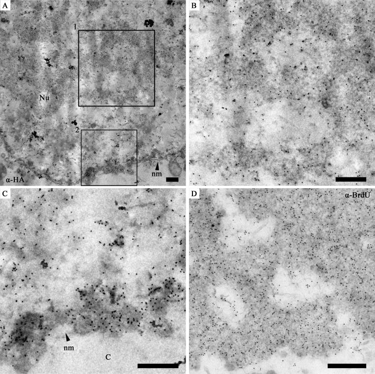
Fig 3.
Immunoelectron micrographs showing the distribution of P6.9 in vP6.9:HA-infected Sf9 cells. (A) A part of a vP6.9:HA-infected cell showing the heavily gold-labeled nucleus. Here, anti-HA was used as the primary antibody, and protein A conjugated with 10 nm gold was used at a 1:50 dilution. (B and C) Enlargement of the vP6.9:HA-infected cell highlighted in panel A. (B) Box 1 from panel A showing gold-labeled P6.9 in the VS. (C) Box 2 from panel A showing that gold-labeled P6.9 localized near the inner nuclear membrane of the nucleus (C). (D) The VS region of a vP6.9HA-infected Sf9 cell. The cell was labeled with BrdU and probed with mouse monoclonal anti-BrdU antibody. Goat anti-mouse secondary antibody conjugated with 10 nm gold was used at a 1:100 dilution. Nu, nucleus; nm, nuclear membrane. Scale bar, 500 nm.

