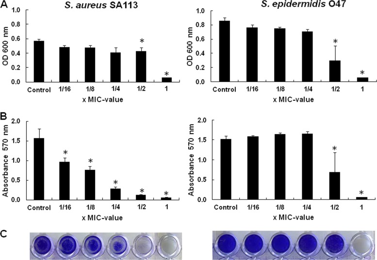
Fig 2.
Biofilm formation of S. aureus SA113 and S. epidermidis O47 in the presence of subinhibitory concentrations of gallidermin. Growth and biofilm formation occurred in microtiter plates. Shown are the turbidity (the OD600) of the cell culture (A), the absorbance (A570) of stained cells attached to the surface after washing (B), and photos of biofilm cells attached to microtiter wells after crystal violet staining (C). Differences were considered significant at P < 0.05.

