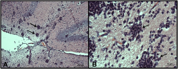
Fig 9.
Light microscopy of olfactory bulbs in mice infected with N. fowleri and treated intraperitoneally with 9 mg/kg/day corifungin for a total of 10 days. (A) Olfactory bulbs show multiple spherical inflammatory foci (arrows) without trophozoites. Some areas of the brain have a discrete vacuolization adjacent to the inflammation, but the parenchyma is well preserved, with no evidence of tissue necrosis. Magnification, ×10. (B) Higher-magnification view of the same zone. Magnification, ×60. The tissues were stained with hematoxylin and eosin.

