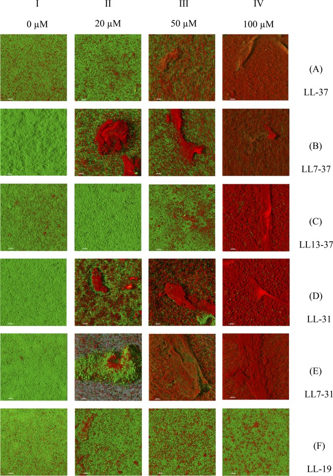
Fig 5.
Effect of six peptides on a GFP-tagged P. aeruginosa PAO1 biofilm stained with PI. A 24-h-old GFP-tagged PAO1 biofilm, grown in the wells of a μClear microtiter plate, was exposed to increasing peptide concentrations in the presence of 10 μM PI for 25 min. After treatment, the biofilm was rinsed and observed by CLSM. Damaged cells are stained red by the entry of PI into the cell, and live cells are stained green by the expression of GFP. The micrograph images represent a tridimensional view from the top of the biofilm and are the most representative of two independent experiments. The white bars on the pictures represent a 20 μM distance.

