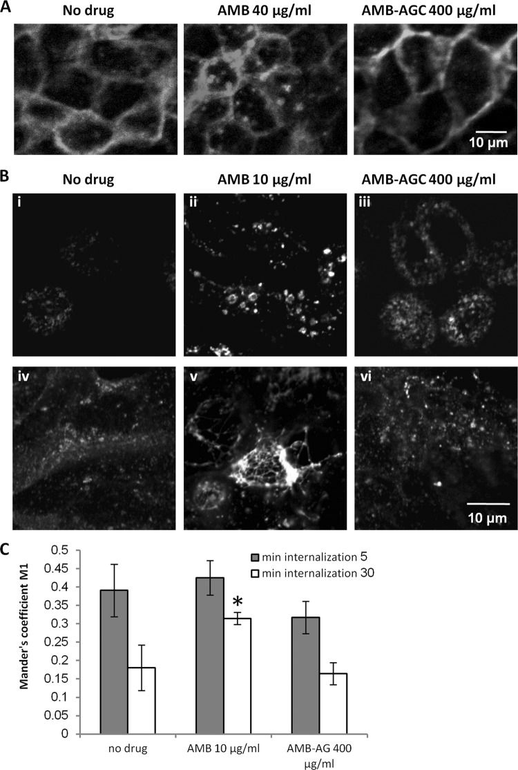
Fig 4.
AMB, but not AMB-AGC, modulates the distribution of receptors in MDCK cells. (A) MDCK cells stably expressing M1-GFP were grown on glass coverslips and treated for 1 h with no drug (control), 40 μg/ml AMB, or 400 μg/ml AMB-AGC. The cells were visualized by fluorescence microscopy after fixation. (B) MDCK-PTR9 cells grown on glass coverslips in MEM/BSA medium were treated for 19 h (i to iii) or 24 h (iv to vi) with AMB (10 μg/ml), AMB-AGC (400 μg/ml), or no drug (control) and then incubated with 60 mg/ml of FITC-hTfn for 60 min on ice to induce binding. After intensive washing, the cells were transferred to fresh medium and incubated at 37°C for 30 min to enable endocytosis. The cells were then washed with acidic buffer to strip membrane-attached FITC-hTfn from the membrane, fixed, and visualized by confocal microscopy. (C) After FITC-hTfn internalization and fixation (19 h treatment), the cells were labeled with mouse anti-EEA1 and then with Cy5-conjugated goat anti-mouse IgG. The samples were documented by confocal microscopy, and quantification of FITC-hTfn colocalization with EEA1 was performed. The Manders' M1 coefficients are presented. Colocalization analyses were performed for at least 20 cells in at least four different microscopic fields. The error bars represent Manders' M1 coefficients ± the SDs. One asterisk represents a P value of 0.00197 compared to the value for the no-drug control. Gray and white columns represent internalization for 5 and 30 min, respectively.

