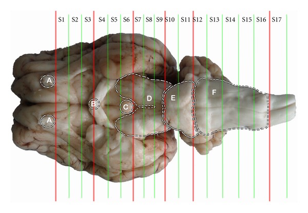
Figure 1.

Brain sectionalization: (A) the residual part of the olfactory bulbs, (B) optic chiasm, (C) mamillary body, (D) cerebral peduncles, (E) Pons and (F) medulla oblongata. In the sections nuclei or areas: nucleus accumbens, septal area and caudate nucleus in S2, supraoptic nucleus, paraventricular nucleus of hypothalamus, and ventromedial nucleus of hypothalamus in S4, arcuate nucleus and amygdala in S5, habenular nucleus in S6, periaqueductal gray in S8, dorsal raphe nucleus and substantia nigra in S9, parabrachial nucleus and locus ceruleus in S10, nucleus raphe magnus in S13, solitary nucleus in S14, gigantocellular reticular nucleus in S15, and spinal cord dorsal horn in S17 are located.
