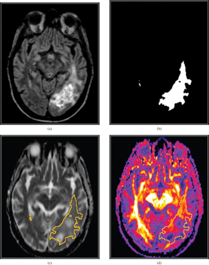
Figure 1.
Image analysis. Three-dimensional (3D) fluid attenuation inversion recovery (FLAIR) image obtained prior to initiation of treatment (a) demonstrates a left parieto-occipital glioblastoma multiforme. The 3D FLAIR image set was segmented to isolate a region of interest defined by FLAIR signal abnormality (FSA). (b) The mean apparent diffusion coefficient and mean fractional anisotropy (FA) were then calculated within FSA from co-registered (c) ADC and (d) FA maps, respectively.

