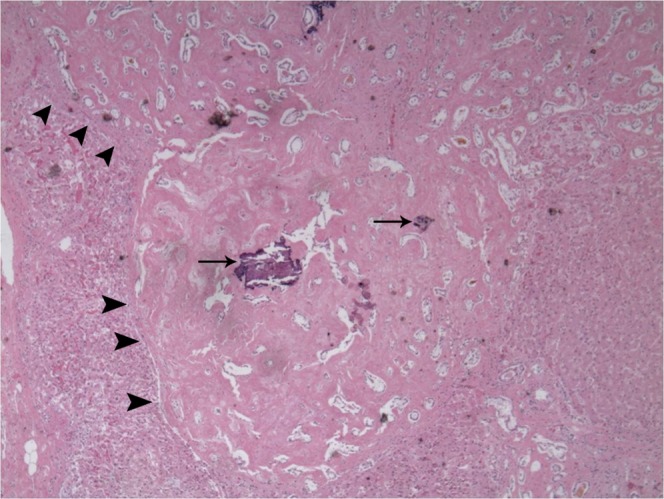
Figure 3.

Haematoxylin and eosin stain of a biliary hamartoma. Nodular areas (arrowheads) with dense hyalinised fibrous stroma and hamartomatous proliferation with irregular ectatic lumens lined with biliary epithelium (original magnification×40). Note the multiple dystrophic calcifications (arrows).
