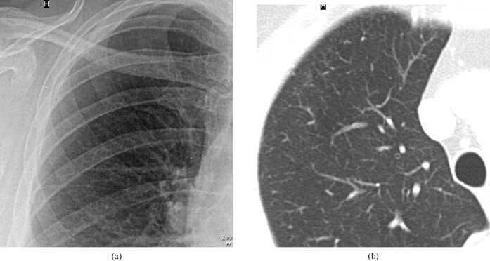
Figure 3.
Correlation of chest radiography and CT appearances in a 69-year-old female smoker (22 pack years). The chest radiograph shows (a) a considerable increase of overall lung markings with small nodular opacities; normal lung markings are still visible. The finding is comparable to the profusion score of small opacities 1/0 according to the International Labour Organization. (b) Transverse CT scan targeted to the right lung shows intralobular thickening and numerous intralobular smooth opacities in the periphery of the upper lobe.

